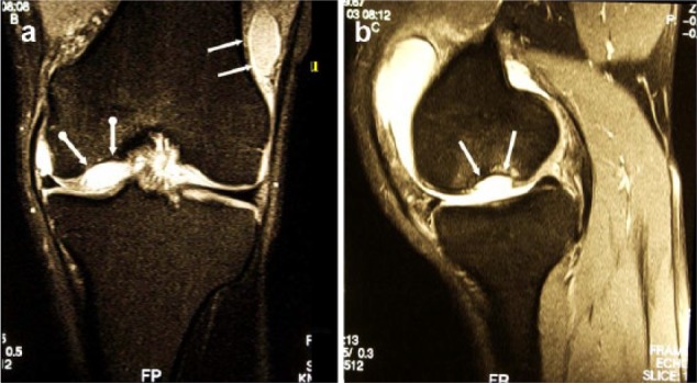
Figure 3.

(a) Magnetic resonance imaging (MRI) (proton density fat saturation) of the same knee joint exhibiting the empty lacuna at the medial condyle (dotted white arrows) and the loose body located at the lateral recessus (white arrows), coronal plane. (b) MRI (proton density fat saturation) of the same knee joint exhibiting the empty lacuna at the medial condyle (dotted white arrows) and the loose body located at the lateral recessus (white arrows), sagittal plane.
