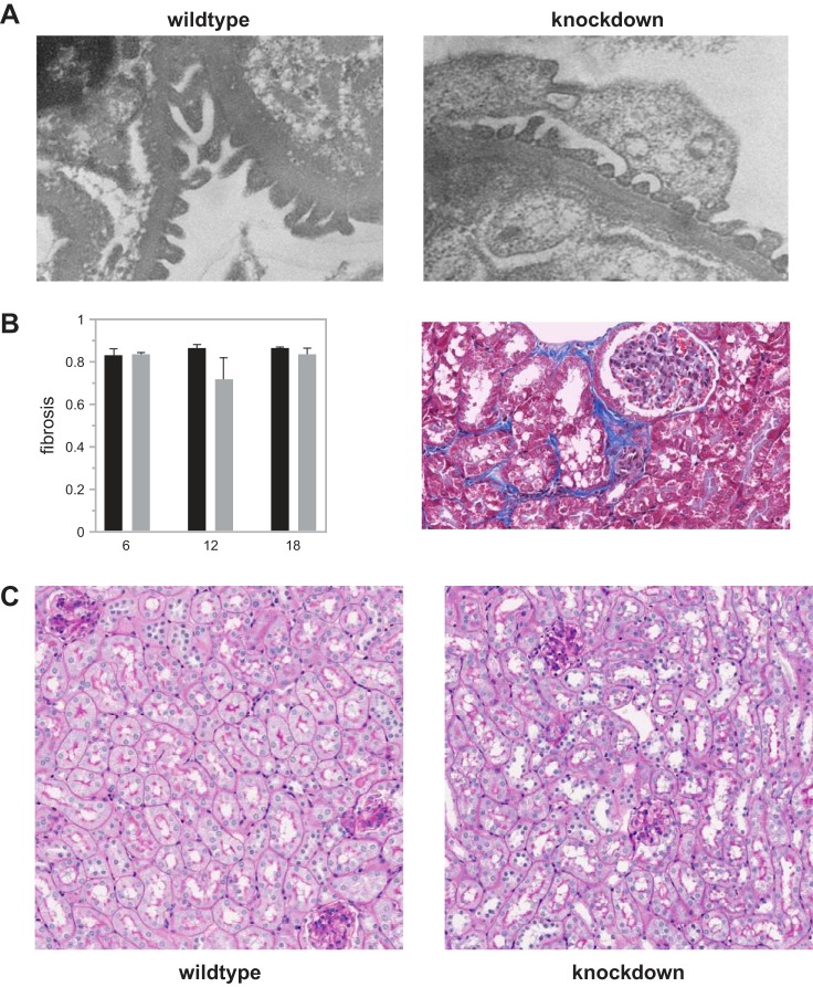
Fig. 7.
Comparison between wild-type and Far2 knockdown animals for podocyte effacement (A), renal fibrosis (B), tubulo-interstitial damage, and immune cell infiltration (C). For podocyte effacement, tubulo-interstitial damage, and immune cell infiltration slides from 10 animals per group and 3 time points were scored independently by two people, and representative pictures from 12 mo old animals are shown. For renal fibrosis we quantified the fibrosis in all animals as described in materials and methods.

