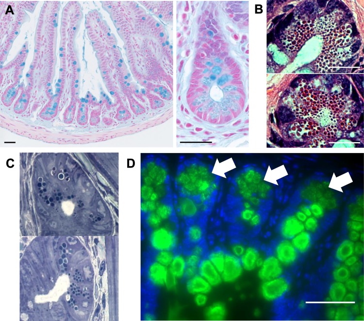
Fig. 1.
C57BL/6 mouse ilea stained with Alcian blue (A). Mucins within the goblet cells and secreted mucus layer are clearly visible. At the crypt bases, Alcian blue staining among the Paneth cell granules is also clearly visible under ×100 and ×400 magnification. Paneth cells at the base of the ileal intestinal crypts of a wild-type C57BL/6 mouse are stained with hematoxylin and eosin (B) or toluidine blue (C) (×1,000 magnification). The granules within the cells are easily visible in the cytoplasm of the cell. Wild-type mouse ileal sections were immunofluorescently stained for Muc2 (green) and DAPI (blue) (D). Strong staining for Muc2 within the goblet cells is clearly visible in green, and fainter fluorescence is visible in the Paneth cells at the base of the crypts (indicated by arrows). Scale bars = 50 μm, ×400 magnification.

