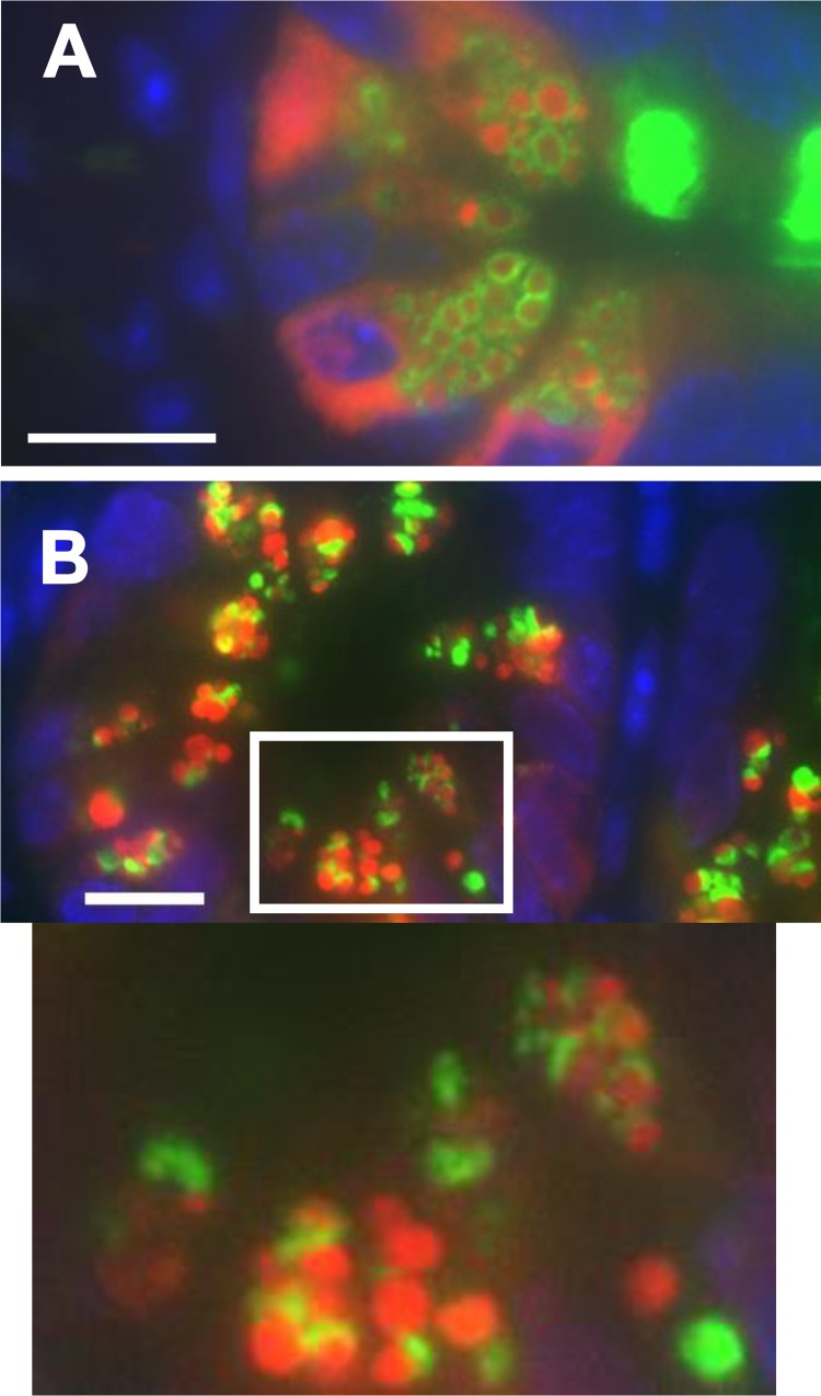
Fig. 5.
Paneth cells from Core-3-deficient (C3Gnt−/−) (A) and Core-1-deficient (C1galt1−/−intestinal epithelial cell) (B) mice, stained for Muc2 (green), lysozyme (red), and DAPI (blue). C3Gnt−/− Paneth cells were indistinguishable from wild-type Paneth cells; however, the C1galt1−/− Paneth cells were lacking most of their Muc2 halo and instead displayed only a patch of Muc2 at 1 pole of each granule (arrows). Granule structure and function were not otherwise affected. Scale bars = 10 μM, ×1,000 magnification.

