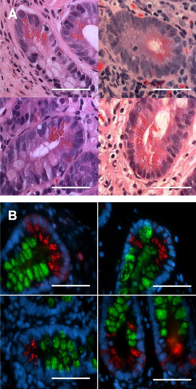
Fig. 8.
Four representative examples of formalin-fixed, hematoxylin and eosin-stained human ileal crypts containing Paneth cells (A). Left: ulcerative colitis. Top, right: patient without inflammatory bowel disease. Bottom, right: patient with Crohn’s disease. No significant differences in Paneth cell morphology were observed between patients, regardless of disease state. Immunofluorescent staining of formalin-fixed human ileal biopsies is shown (B). MUC2 is represented in green, lysozyme in red, and the nuclei are stained for DAPI in blue. The 4 panels are at ×1,000 magnification of the base of the crypts, where the Paneth cells reside. MUC2 staining within the epithelium is largely limited to the goblet cells lining the villi and crypts and is also visible within the thick mucus layer lining the tissue. The lysozyme is largely limited to the Paneth cells as the base of the crypts, and, unlike the murine Paneth cells, there were no signs of positive MUC2 staining within human Paneth cells. Scale bars = 20 μM, ×630 magnification.

