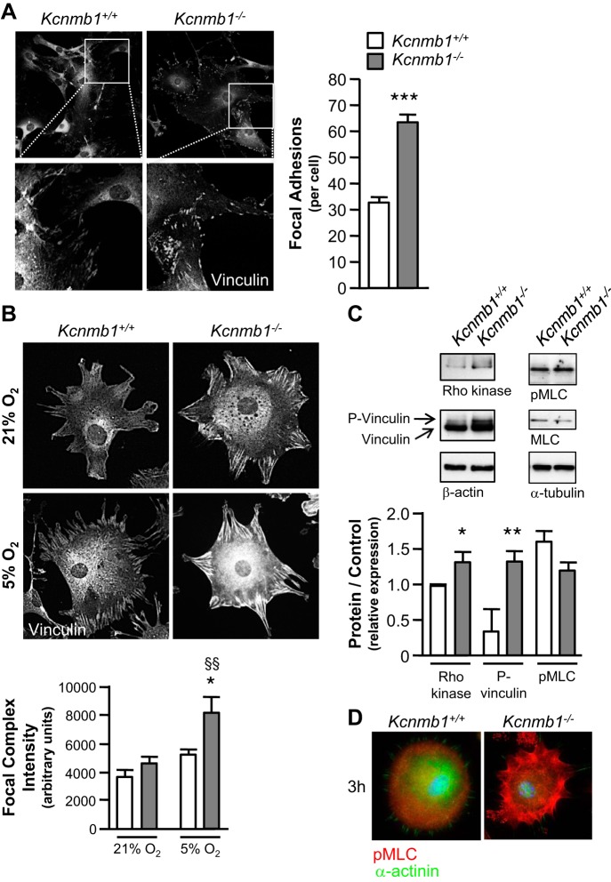
Fig. 6.
Hypoxia induces greater focal complex expression in Kcnmb1−/− compared with Kcnmb1+/+ pulmonary artery smooth muscle cells (PASMCs). A: expression of focal adhesions in isolated peripheral PASMCs from Kcnmb1+/+ and Kcnmb1−/− mice. Quantification of the number of focal adhesions per cell 24 h postseeding as visualized by vinculin expression, magnification ×200. Bars represent means ± SE (n = 3). ***P ≤ 0.001, Kcnmb1−/− vs. Kcnmb1+/+. B: focal complex expression in peripheral PASMCs. Hypoxia (5% O2) increases focal complexes to a greater degree in Kcnmb1−/− compared with Kcnmb1+/+ PASMCs. Quantification of focal complex intensity per cell (arbitrary units) 3 h postseeding as visualized by vinculin expression, magnification ×200. Bars represent means ± SE (n = 3). *P ≤ 0.05, 5% O2 Kcnmb1−/− vs. 5% O2 Kcnmb1+/+; §§P ≤ 0.01, 5% O2 Kcnmb1−/− vs. 21% O2 Kcnmb1−/−. C: Western immunoblots of Rho kinase, vinculin, phospho-vinculin (P-vinculin), β-actin, phospho- myosin light chain (pMLC), MLC, and α-tubulin expression in isolated PASMC. Rho kinase and phospho-vinculin expression are increased in PASMC from Kcnmb1−/− compared with Kcnmb1+/+ 3 h postseeding. Bars represent means ± SE (n = 3). *P ≤ 0.06, **P ≤ 0.01, Kcnmb1−/− vs. Kcnmb1+/+. D: representative images of the expression of pMLC in isolated peripheral PASMC 3 h postseeding. pMLC, red; α-actinin, green; nuclei, blue; magnification ×400.

