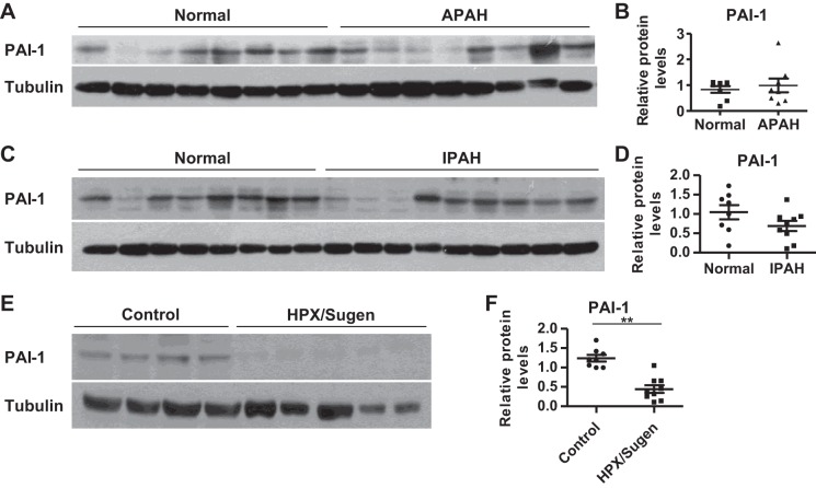
Fig. 7.
Expression levels of PAI-1 in pulmonary arterial hypertension (PAH) patients or when exposed to hypoxia or hypoxia mimics. A and B: PAI-1 protein levels in human PASMCs of associated (A)PAH patients and normal donors were determined and compared. One normal donor’s PAI-1 level was set as 1 and was used for normalization for that of other normal donors and APAH patients; 8 normal donors and 8 APAH samples. C and D: PAI-1 protein levels in human PASMC of idiopathic (I)PAH patients and normal donors were determined and compared. One normal donor’s PAI-1 level was set as 1 and was used for normalization for that of other normal donors and IPAH patients; 8 normal donors and 9 IPAH samples. E and F: PAI-1 protein levels in lungs of control rats and hypoxia/Sugen-treated rats (HPX/Sugen) were determined and compared. One control rat lung’s PAI-1 level was set as 1 and was used for normalization for that of other control and HPX/Sugen rat lungs; 8 control rats and 9 HPX/Sugen-treated rats. Representative blots and quantification are presented in B, D, and F. Data are presented as means ± SE. *P ≤ 0.05, **P ≤ 0.01.

