Figure 1.
Localization and Lys Acetylation Activity of NSI.
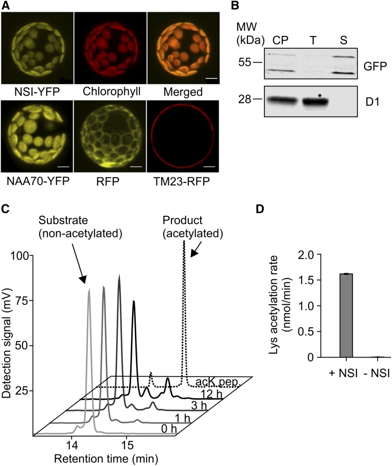
(A) Confocal microscopy image of Arabidopsis protoplast transiently expressing NSI-YFP (35S:NSI-YFP) fusion protein. Upper left panel shows the YFP signal, middle panel chlorophyll fluorescence of the same protoplast, and right panel is a merged image of the two. The lower panel shows the control lines: NAA70-YFP (left) was used as a chloroplast control marker, RFP (middle) as a cytoplasmic control, and TM23-RFP (right) as a plasma membrane control. Bar = 10 µm.
(B) Immunoblot detection of chloroplast protein fractions isolated from transgenic plants expressing NSI-YFP (35S:NSI-YFP) and separated on 12% acrylamide gel. GFP antibody was used for the detection of NSI-YFP and D1 antibody as a thylakoid membrane marker. NSI-YFP was detected as two bands, which may represent the preprotein (molecular weight based on mobility = 61.0 kD; expected molecular weight = 56.5 kD) and processed mature protein (molecular weight based on mobility = 49.0 kD; expected molecular weight = 49.9 kD). Ten micrograms of protein was loaded per sample. CP, chloroplasts; T, thylakoid fraction; S, soluble fraction.
(C) HPLC analysis of a general lysine acetyltransferase substrate and its acetylated product after conversion by His6-NSI for 1, 3, or 12 h. Identities of nonacetylated (0 h) and acetylated (acK pep.) standard peptides were confirmed by MS.
(D) Lysine acetylation rate of a peptide substrate by 10 µM His6-NSI (n = 3 technical replicates, ±sd).

