Figure 3.
Organization of Thylakoid Protein Complexes of the Wild Type, nsi, and stn7 and Thylakoid Ultrastructure of the Wild Type and nsi.
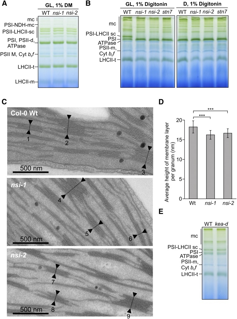
(A) Large-pore blue native gel of thylakoid protein complexes from thylakoids that were isolated from GL (100 µmol photons m−2 s−1) adapted plants and solubilized with 1% β-dodecylmaltoside (DM). Representative image from experiment repeated with three biological replicates is shown. mc, megacomplex; sc, supercomplex; t, trimer; d, dimer; m, monomer.
(B) Large-pore blue native gels after digitonin solubilization of thylakoids isolated from plants after GL or D adaptation.
(C) Transmission electron microscopy (TEM) analysis of the nsi chloroplasts. TEM pictures of palisade mesophyll cells with chloroplasts in close-up view. Leaves of the two T-DNA insertion lines nsi-1 and nsi-2 and of wild-type Col-0 were prepared as thin section samples. Numbers and arrows display exemplary thylakoid stacks.
(D) Average heights per granum membrane layer ± sd for the two nsi knockout lines in comparison to the wild type Col-0 (calculated from [C]). Seven hundred thylakoid stacks per plant line displayed in 70 TEM pictures from seven independent biological replicates were analyzed (***P ≤ 0.001 using two-tailed Student’s t test).
(E) Large-pore blue native gel of GL adapted wild-type and kea1 kea2 double knockout (kea-d) thylakoids solubilized with 1% digitonin.

