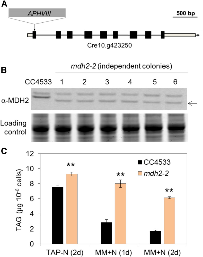
Figure 3.
mdh2-2 Mutant Characterization.
(A) The insertion site of the cassette APHVIII in mdh2-2.
(B) Immunoblot analysis using anti-MDH2 antibodies.
(C) TAG content analysis.
Cells were grown in mixotrophic conditions under constant light. TAG contents were determined in N-deprived cells (2 d) and in N-resupplied cells (1d and 2d). Values are the mean of biological replicates (i.e., independent shaking flask cultures; n = 3, sd). Asterisks indicate statistically significant changes compared with parental strain (CC4533) by paired-sample Student’s t test (**P ≤ 0.01). Note: We have screened six independent lines, and all of them possessed a hybridizing signal just below the expected size, suggesting the likely formation of a truncated protein (black arrow). For immunoblot, samples were loaded at equal total protein amounts. Loading controls were stained by Coomassie blue.

