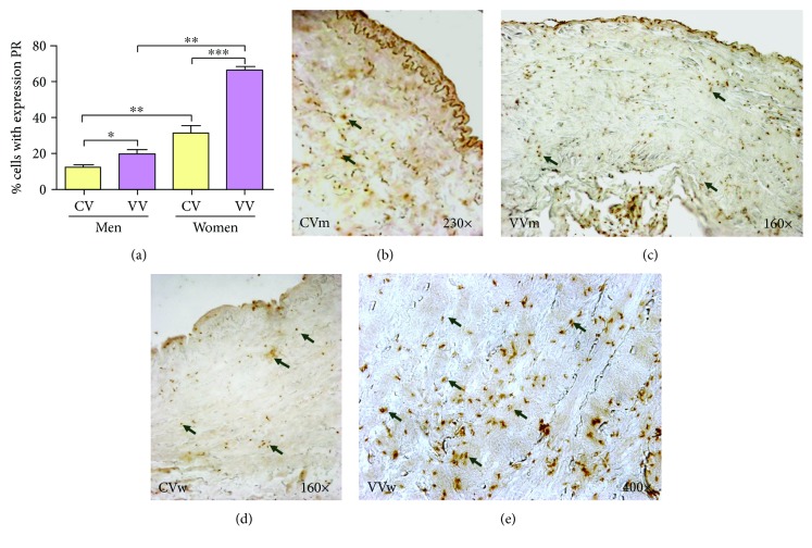
Figure 2.
(a). Percentage of cells positively expressing the progesterone receptor (PR) in the study groups (CV = control vein, VV = varicose vein). (b–e). PR immunodetection images from the four analysis groups. The brown colour indicates the precipitate that correlates with PR protein expression. ∗p < 0.05, ∗∗p < 0.01, and ∗∗∗p < 0.001.

