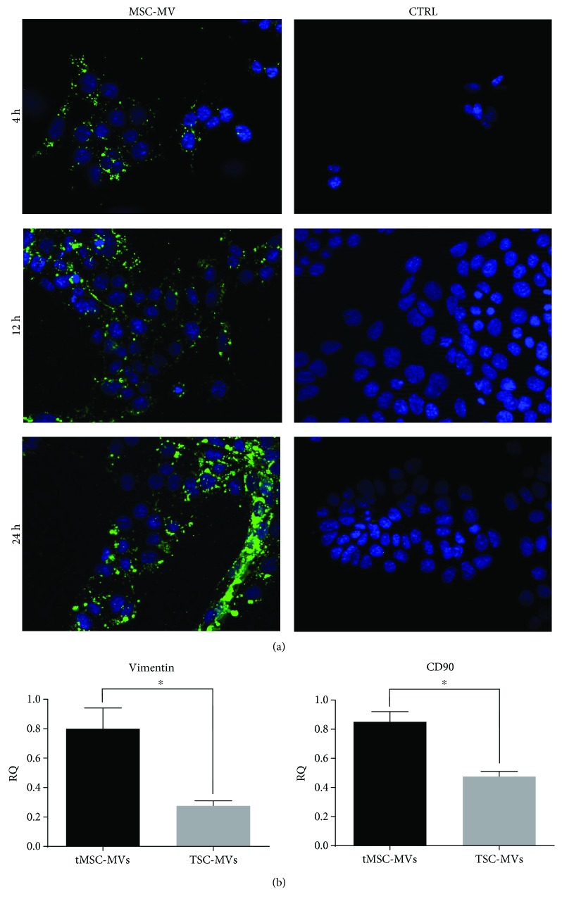
Figure 4.
GFP+ MVs derived from tMSCs are incorporated by IMCD3. (a) Representative pictures of IMDC3 incubated with GFP-labelled MVs at 4, 12, and 24 hours and the control (CTRL) cells incubated only with the labelling reagent. The MVs are progressively fused with the cells as demonstrated by the increased amount of fluorescence. Original magnification: ×400. (b) Finally, real-time analysis shows that MVs derived from tMSCs are positively enriched in vimentin and CD90 when compared to the MVs obtained from testicular somatic cells isolated from the same biopsy (TSC-MVs) (N = 3, ∗ P < 0.05).

