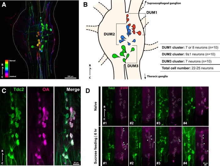
Figure 7.
Sucrose feeding-evoked reporter protein expression in the DUM neurons of the IEG reporter line. A, Dorsal view of the subesophageal ganglion of the Hokudai WT strain stained with anti-Gryllus Tdc2 antibody. The outline of the ganglion is surrounded by the white dotted line. The depth of the cells is color coded as indicated in the inset. Rostrocaudal (R-C) axis was indicated. Scale bar represents 200 µm. See Fig. 7-1 for the octopamine biosynthesis pathway and the structures of the Tdc proteins in insects. See Fig. 7-2 for the frontal view of the supraesophageal ganglion and the ventral view of the subesophageal ganglion stained with anti-Gryllus Tdc2 antibody. B, Schematic drawing of the positions and numbers of the cell bodies of three DUM clusters (DUM1, DUM2, DUM3) on the dorsal side of the subesophageal ganglion. C, Double fluorescent immunostaining confirmed that the DUM neurons contain octopamine. The DUM neurons were stained with the anti-Gryllus Tdc2 antibody (green) and anti-octopamine antibody (magenta). Scale bar represents 50 µm. D, Distribution of the reporter protein (EYFPnls:PEST) in the DUM neurons of the IEG reporter line before and 6 h after feeding of sucrose solution (n = 4 each). The DUM neurons were stained with the anti-Gryllus Tdc2 antibody (green) and anti-GFP antibody (magenta). The cell bodies of Gryllus Tdc2 immunoreactive DUM neurons are surrounded by the white dotted line. The DUM neurons with nuclear EYFP immunoreactivity are indicated by white arrowheads. Scale bar represents 50 µm.

