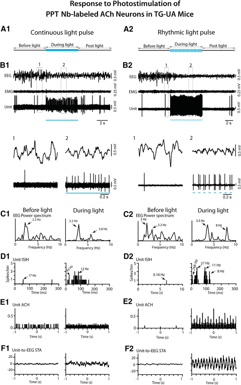
Figure 11.
Response of PPT Nb-labeled ACh units to continuous and rhythmic light pulse stimulation in TG-UA mice. A1, A long (∼5 s) continuous light pulse was delivered during a period of EEG irregular slow wave activity. B1, The unit (#14 in mouse CChAT13) markedly increased its discharge while the EEG concomitantly shifted from irregular slow wave activity to more rhythmic θ activity, as shown in expanded segments (1, 2 below). C1, In the EEG power spectra, a shift in peak frequencies from 2.2 to 3.2 and 5.8 Hz was apparent. D1, In the unit ISIH, the increased discharge by the unit appeared to consist of irregular tonic firing (mode of 12 Hz indicated by arrow) with some phasic activity (∼43 Hz, arrow). E1, In the unit ACH, there was no evidence of rhythmic firing. F1, In the unit-to-EEG STA, there was no evidence of cross-correlated activity. A2, Rhythmic light pulse stimulation at high θ frequency (50 ms, 8 Hz for ∼5 s) was delivered during a period of EEG irregular slow wave activity. B2, The unit (#5 in mouse CChAT11) was driven to fire in a phasic manner with bursts of spikes with each light pulse, and the EEG was concomitantly driven to rhythmic θ oscillations, as shown in expanded traces (1, 2 below). C2, In the EEG power spectra, the activity appeared to shift from mixed irregular slow wave activity to more rhythmic activity with peaks at 3.5 and 8 Hz. D2, In the unit ISIH, the firing appeared to consist of phasic activity of clusters (with mode at 27 Hz, indicated by arrow) and some bursts (166 Hz, arrow) of spikes and tonic firing (mode at 11 Hz, arrow) along with the recurrent θ rhythm (mode at 8 Hz, arrow). E2, In the unit ACH, a degree of rhythmicity in firing at around 8 Hz was apparent. F2, In the unit-to-EEG STA, evidence of cross-correlated discharge of the unit with EEG θ activity 8 Hz was apparent. See legend to Figure 5 for graph details and abbreviations.

