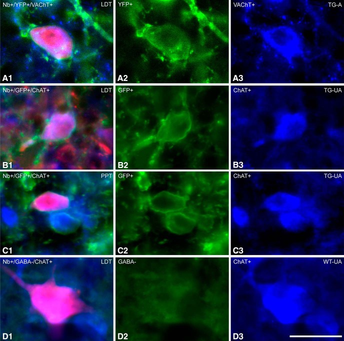
Figure 2.
Fluorescent images of ACh recorded and Nb-labeled cells. Neurons which were recorded and labeled with Nb (in red) were identified as ACh neurons by immunofluorescent staining for VAChT or ChAT (in blue). A, Located in the LDT of a TG-A mouse (ChAT8), an Nb-labeled cell, which was VAChT+, appeared to express ChR2-EYFP (evident as YFP over the plasma membrane). B, Located in the LDT of a TG-UA mouse (Chronic (C)ChAT8), an Nb-labeled cell, which was ChAT+, appeared to express ChR2-EYFP (revealed by immunohistochemical staining for GFP over the plasma membrane). C, Located in the PPT of a TG-UA mouse (CChAT13), an Nb-labeled cell, which was ChAT+, also appeared to express ChR2-EYFP (revealed by immunohistochemical staining for GFP over the plasma membrane). D, Located in the LDT of a WT-UA mouse (CWT8), an Nb-labeled cell, which was ChAT+ was also confirmed to be immunonegative for GABA.

