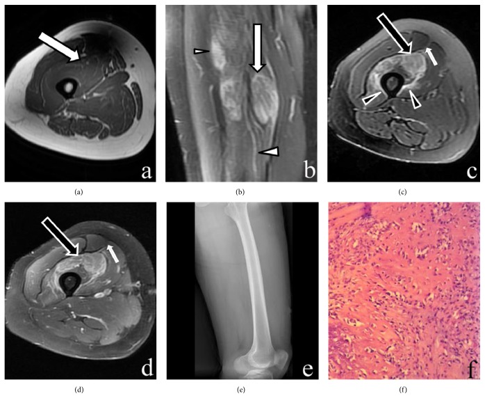
Figure 3.
MR, DR, and histopathology imaging of the right thigh. (a) Axial T1-weighted image shows an ill-defined isointense lesion in the vastus intermedius muscle (large arrow). (b) Coronal fat-suppressed T2-weighted image reveals a hyperintense lesion with a “striate pattern” in the vastus intermedius muscle (large arrow). (c) Axial fat-suppressed T2-weighted image shows a hyperintense lesion with a “checkerboard-like pattern” in the vastus intermedius muscle (large arrow). Surrounding soft-tissue edema in the vastus intermedius and vastus medialis muscles (arrowheads) and overlying fascia (small arrow) is seen. (d) Enhanced fat-suppressed axial T1-weighted image shows that the lesion enhances intensely with a “checkerboard-like pattern” (large arrow). Preservation of the muscle fascicles is shown within the lesion. Enhancement of the overlying fascia (small arrow) is seen. (e) Lateral DR shows the anterior femoral soft tissue without any calcification or ossification. (f) The specimen mainly includes loose, immature textured fibroblasts with mild cellular pleomorphism. Some portions of the lesion contained osteoid formation. The entrapped atrophic or necrotic muscle fibers are also shown in the lesion (H&E staining, ×200).

