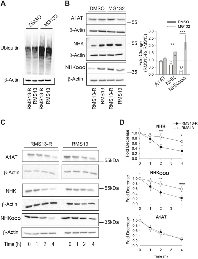
Fig. 1.
RMS13-R cells exhibit heightened levels of ERAD. (A) The MAL3-101-resistant rhabdomyosarcoma cell line (RMS13-R) and the parental MAL3-101-sensitive cell line (RMS13) were treated with DMSO or 20 µM MG132 for 4 h followed by an immunoblot analysis to visualize the total ubiquitylated protein. In the presence of MG132, there was a 2.5-fold increase in ubiquitylated protein levels. (B) Relative ERAD efficiency was measured by analyzing the steady-state levels of the NHK and NHKQQQ mutants in the presence or absence of 20 µM MG132 for 4 h. A1AT was used as a negative control. The graph on the right shows the ratio between the levels of each protein in the RMS13-R and RMS13 cells after treatment with DMSO (in white) and MG132 (in gray). The mean±s.d. of eight different experiments are plotted. (C,D) Both NHK and NHKQQQ are degraded faster in RMS13-R cells. The RMS13-R and RMS13 cells were treated with cycloheximide after a 3-h MG132 pre-treatment, aliquots were removed at the indicated times and lysates were resolved by SDS-PAGE and immunoblotted to detect A1AT abundance. Graphs represent the mean±s.d. of four independent experiments. *P<0.05, **P<0.005, ***P<0.0001.

