Fig. 3.
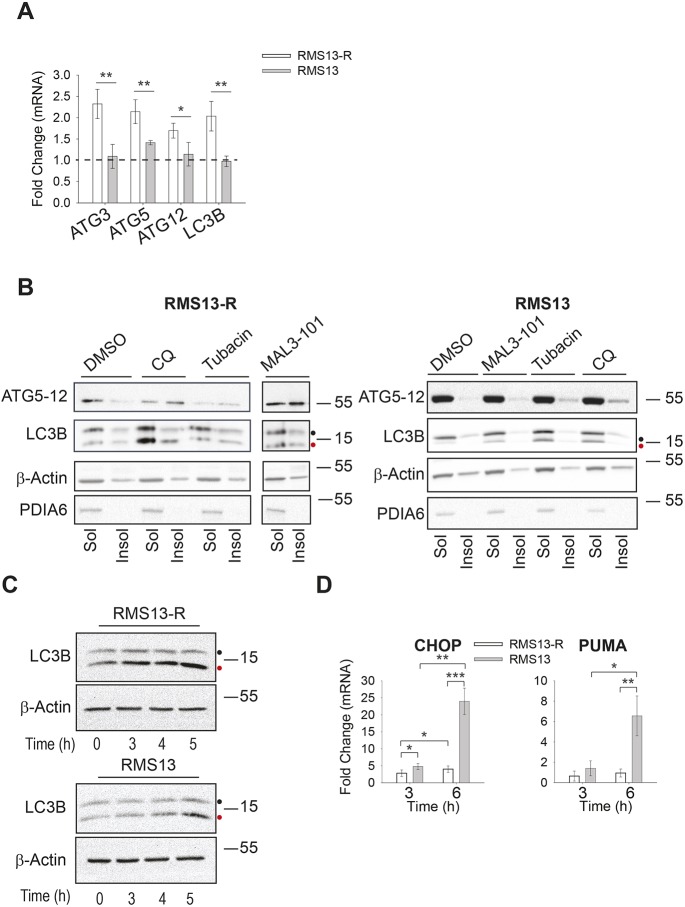
Hsp70 inhibition induces autophagy in RMS13-R cells. (A) The mean±s.d. relative level of autophagy gene expression was detected by qPCR in the presence of DMSO or 7.5 µM MAL3-101 for 6 h in RMS13-R (white) and RMS13 (gray) cells (n=4). *P<0.05, **P<0.005. (B) The amount of the indicated soluble (Sol) and membrane-associated (Insol) proteins was examined by immunoblotting after treating cells with 50 µM CQ, 11 µM tubacin or 7.5 µM MAL3-101 for 6 h. PDIA6, a soluble ER-resident chaperone, was used as a control. (C) RMS13-R and RMS13 cells were treated for the indicated times with 7.5 µM MAL3-101 and CQ was added during the last hour of the treatment to test autophagic flux. Therefore, time ‘0’ indicates a 4 h treatment with DMSO plus a 1 h treatment with CQ. Aliquots of cell lysates were resolved by SDS-PAGE and immunoblotted for LC3B as a marker for autophagic flux. LC3BI is indicated by a black dot and the autophagosome-associated form LC3BII is highlighted with a red dot. (D) RMS13-R or RMS13 cells were treated for 3 or 6 h with 7.5 µM MAL3-101 and the expression of the pro-apoptotic CHOP and PUMA genes was measure by qPCR relative to the DMSO control as the mean±s.d. (n=3). *P<0.05, **P<0.001, ***P<0.0001.

