Fig. 5.

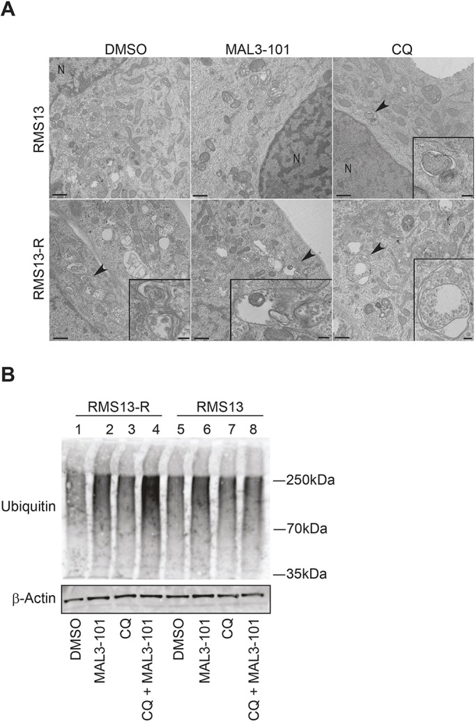
RMS13-R cells show evidence of constitutive autophagy. (A) Electron microscopy analysis was conducted on RMS13 and RMS13-R cells treated with DMSO, 7.5 µM MAL3-101, or 50 µM CQ for 4 h. Insets (areas indicated by black arrowheads) under high magnification are shown in the bottom right and provide evidence of autophagosomes or autophagic structures. Of note, heterochromatin in the nucleus (indicated by ‘N’) as well as disrupted mitochondrial morphology are visible upon MAL3-101 treatment in RMS13 cells. Scale bars: 200 nm. (B) RMS13-R and RMS13 cells were treated with DMSO, MAL3-101, CQ or MAL3-101/CQ for 4-h, lysates were prepared, and cellular protein was immunoblotted for total ubiquitylated proteins. When CQ was administrated together with MAL3-101 to RMS13-R cells, ubiquitylated protein content increased ∼3-fold compared to the DMSO control.
