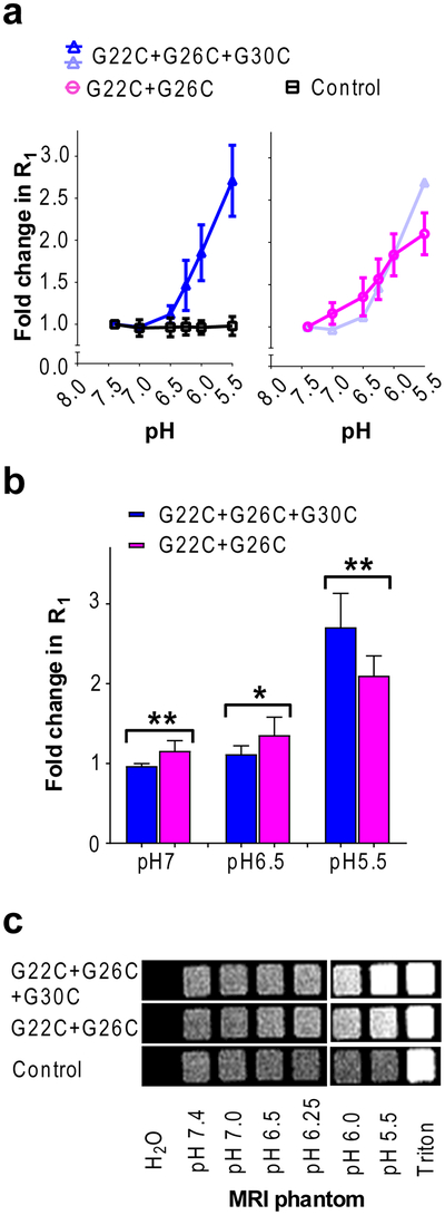
Figure 4: pH dependent R1 increase and MR contrast enhancement of Gd-DOTA liposomes reconstituted with modified nanopores.
(a). The R1 relaxation rates at 1.4 T and 25 °C. (b). Comparison of R1 for the double and triple labeled mutant. “**”, p < 0.01, t = 3.83, df = 7; “*” p < 0.05, t = 2.48, df = 10; “**”, p < 0.01, t = 3.28, df = 12; student t test. (c). Spin echo T1 weighted MR image (at 1 T) of phantoms. Imaging parameters: TR = 200 ms, TE = 13.1 ms, data matrix = 128 × 128, FOV = 70 mm, slice thickness = 1 mm.

