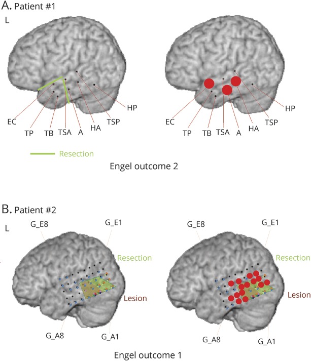
Figure 4. Two patient examples from the Freiburg cohort.
In both patients, not all high-frequency oscillations (HFOs) were removed. (A) In patient 1, mesial temporal contacts in the hippocampus (HA) and amygdala (A) had HFOs in the deepest contacts and were not removed because the patient had a pole resection, including an encephalocele, in this area and the mesial temporal structures were spared. The patient had Engel 2 outcome. Therefore, the prognostication was judged to be correct. (B) The second patient had a lesionectomy (brown) as shown. Red dots indicate contacts with highest fast ripple rates. Not all of these contacts were removed, as shown in the resection line (green). The patient was seizure-free after surgery; therefore, HFOs failed to predict outcome. EC = encephalocele; G = gyrus; HP = parahippocampal gyrus; TB = temporobasal; TP = temporopolar; TSA = temporalis superior anterior; TSP = temporalis superior posterior.

