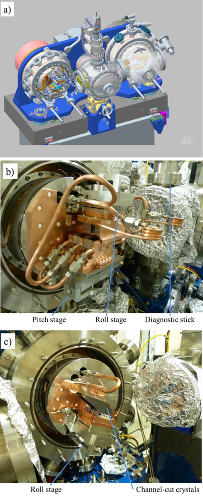
Figure 4.
(a) Mechanical layout of the four-crystal monochromator. The direction of the beam is from left to right in the figure. (b) Photograph of the upstream axis showing the crystal cage fully assembled; an insertion diode and fluorescence screen are mounted on the diagnostic stick. (c) Photograph of the downstream axis showing the channel-cut crystals.

