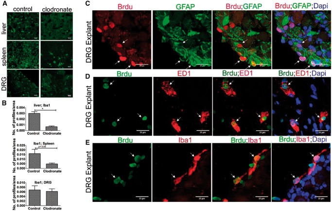
FIGURE 4.
Blood monocytes deprivation has no effect on proliferation of resident macrophages and SGCs in DRG. (A) Clodronate-liposome treatment completely or partially depletes macrophages (green; Iba1+) in liver and spleen respectively, but not DRG. Scale bar: 25 µm. (B) Quantification of (A) shows depletion of Iba1+ macrophages in the liver and spleen, but not DRG, after clodronate treatment. Iba1+ profiles were measured using Image J threshold method and expressed as number of profiles/area, n = 2/group. For liver, p = 0.0209, df = 2, *p < 0.05 (Student t-test, two-tailed); spleen, p = 0.0698, df = 2 (Student t-test, one-tailed), DRG, p = 0.4015, df = 2 (Student t-test, one-tailed). (C) Brdu uptake in GFAP+ cells in the DRG explant indicates local turnover of SGCs. (D) Brdu uptake in ED1+ cells in the DRG explant indicates local turnover of macrophages. (E) Brdu uptake in Iba1+ cells in the DRG explant indicates local turnover of macrophages. (Brdu+ cells expressing GFAP, ED1 or Iba1 are shown using arrows). Scale bar: 25 µm.

