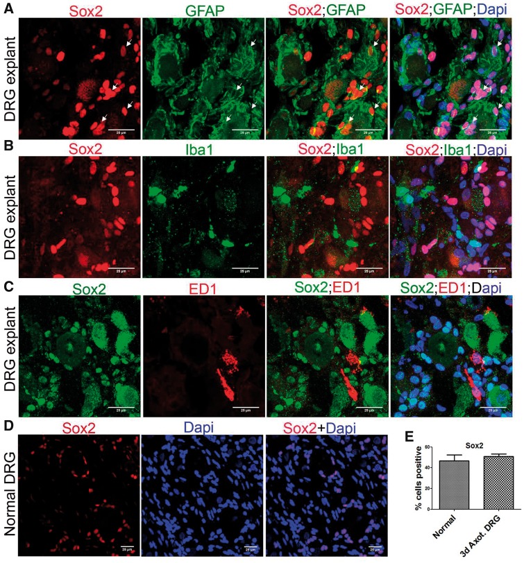
FIGURE 7.
SGCs selectively express Sox2 in DRG explants. (A) Co-expression of Sox2 and GFAP in DRG explant shows Sox2+ SGCs (GFAP+/Sox2+ cells are shown using arrows). (B) Expression of Sox2 and Iba1 in DRG explant shows lack of Sox2 expression in Iba1+ macrophages. (C) Expression of Sox2 and ED1 in DRG explant shows lack of Sox2 expression in ED1+ macrophages. (D) Sox2+ cells are present in adult uninjured DRG. (E) Quantification of number of Sox2+ cells in normal and 3-day axotomized DRGs shows no significant difference. The number of Sox2+ cells is expressed as the percentage of total number of DAPI+ cells, n = 3 animals for each group, number of cells, 1491 for normal, 1915 for axotomized DRG, no statistical significance (Student t-test, two-tailed). Scale bar: 25 µm.

