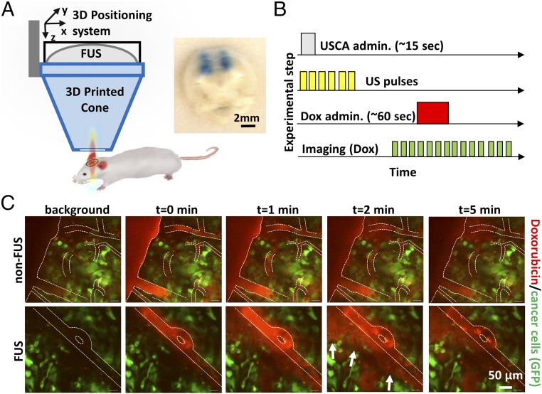
Fig. 1.
FUS enhances doxorubicin extravasation in BT474-Gluc brain tumors. (A) FUS system and experimental setup. (Inset) Image of trypan blue extravasation in gross pathology of a coronal plane section after FUS-BTB disruption in healthy mice (480-kPa peak negative pressure). (B) Schematic illustration of the drug administration protocol. Ultrasound contrast agent (USCA–Definity; Lantheus Medical Imaging) was administered as a bolus. (C) Representative sequential images from intravital multiphoton microscopy of doxorubicin distribution in the breast cancer BM model with (Lower) and without (Upper) FUS-BTB disruption. Red, doxorubicin autofluorescence; green, GFP-positive BT474-Gluc cancer cells.

