Fig. 1.
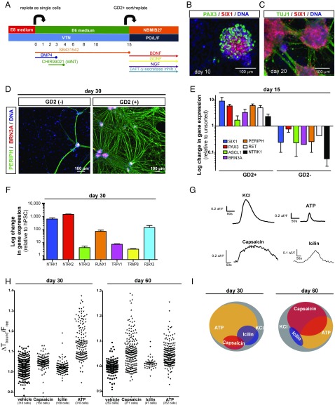
Differentiation and functional characterization of trigeminal neurons derived from human iPSC under fully defined conditions. (A) Modified trigeminal placode induction protocol using only fully defined components. VTN, vitronectin. (B) Placodal clusters staining for SIX1 (cranial placode) and PAX3 (trigeminal placode) after 10 d of differentiation. (C) Placodal clusters rapidly differentiate into TG neurons staining positive for TUJ1 and SIX1 after 20 d of differentiation. (D) Sorting for GD2 on day 15 of differentiation results in highly pure TG neurons staining for peripherin and Brn3a after 15 additional days of differentiation. (E) Gene-expression analysis of key sensory and TG neuron markers after GD2 sorting on day 15 of differentiation. Data are expressed as fold-changes compared with unsorted cells. (F) Gene-expression analysis of nociceptor genes on day 30 of differentiation. (G) Representative individual calcium traces for the four different stimuli used. (H) Calcium response to capsaicin, icilin, and ATP of each individual cell analyzed on day 30 and day 60 of differentiation. (I) Venn diagram showing subgroups of TG neurons that respond to KCl, capsaicin, icilin, and ATP.

