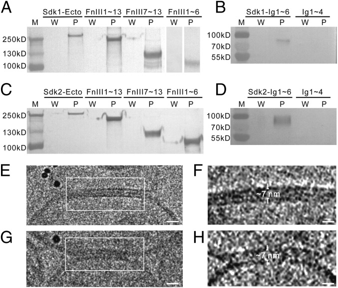
Fig. 3.
The FnIII domains of Sdks interact with liposomes. The supernatants after washing (W) and the liposome pellets (P) are assayed by Western blot (A–D). (A) The ectodomain and the FnIII domain-only fragments (Sdk1 FnIII1∼13, Sdk1 FnIII1∼6, Sdk1 FnIII7∼13) of Sdk1 can be pulled down by liposomes. (B) Sdk1 Ig1∼6 can be pulled down by liposomes, while Sdk1 Ig1∼4 has no interaction with liposomes. (C) The ectodomain and the FnIII domain-only fragments (Sdk2 FnIII1∼13, Sdk2 FnIII1∼6, Sdk2 FnIII7∼13) of Sdk2 can be pulled down by liposomes. (D) Sdk2 Ig1∼6 can be pulled down by liposomes, while the Sdk2 Ig1∼4 has no interaction with liposomes. (E and G) Cryo-images of adhesion interfaces (white square) formed between the Sdk1 ectodomain-incorporated liposomes. (F and H) Tomographic slices of the adhesion interfaces indicated in E and G, respectively. The intermembrane distances are indicated and labeled. (Scale bars: E and G, 15 nm; F and H, 10 nm.)

