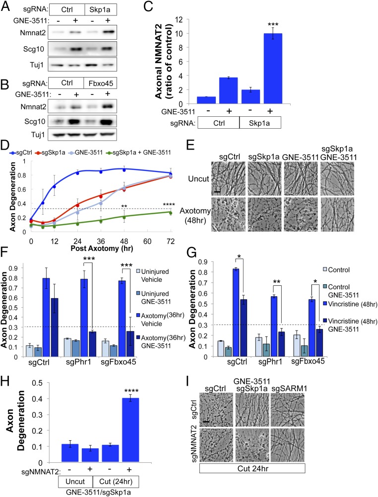
Fig. 6.
Combined inactivation of MAPK signaling and E3 ligase complex confers additive axon protection. Cas9-expressing DRG sensory neurons transduced with control (Ctrl) sgRNAs or sgRNAs targeting Skp1a (A) or Fbxo45 (B) were treated with GNE-3511 for 14 h, and levels of endogenous NMNAT2 were assessed from axon-only extracts by Western blot. (C) Quantification of axonal NMNAT2 levels from DRGs treated as in A [***P < 0.001, single-factor ANOVA with Bonferroni post hoc test for multiple comparisons (n = 3)]. (D and E) Time course of axon degeneration after axotomy from DRGs treated with vehicle or GNE-3511 and Ctrl sgRNAs or sgRNAs targeting Skp1a. Axons are preserved for 72 h after axotomy when pretreated with GNE-3511 and sgRNAs to Skp1a, with representative images shown in E. (Scale bar, 5 μm.) A repeated-measures ANOVA was performed with Bonferroni post hoc tests for multiple comparisons, where ****P < 0.0001 and **P < 0.005. (F) Axon degeneration in distal axons 36 h after axotomy from DRGs treated with DMSO or GNE-3511 and sgRNAs targeting Phr1 or Fbxo5. (G) Axon degeneration in distal axons from DRG sensory neurons treated with 40 nM vincristine transduced with the indicated sgRNAs and GNE-3511. For F and G, a two-way ANOVA with post hoc Bonferroni test for multiple comparisons was performed, where *P < 0.01, **P < 0.005, and ***P < 0.001 (n = 3). (H) Cas9-expressing DRGs were treated as described in D, except cells were transduced with sgRNAs to NMNAT2 on DIV4. Axon degeneration was assessed in severed axons 24 h after axotomy (two-way ANOVA with Bonferroni correction for multiple comparisons, where ****P < 0.0001). Error bars represent SEM. (I) Representative bright-field images of distal axons from H with sgRNA to SARM1 included as a control. (Scale bar, 5 μm.)

