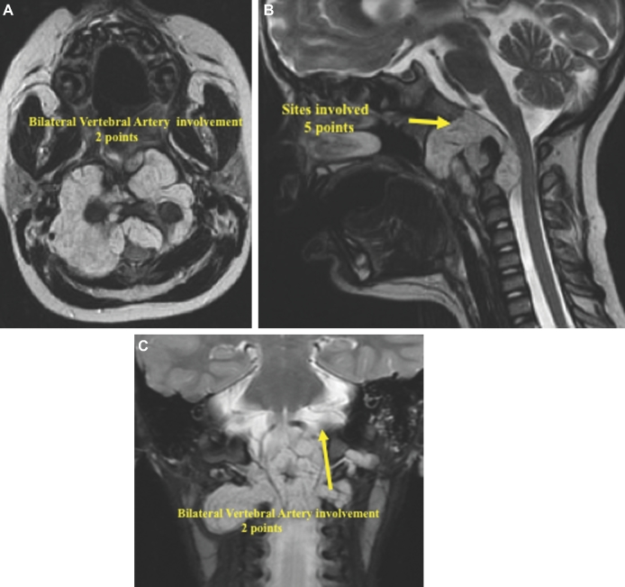
FIGURE 8.
A-C, The preoperative MRI imaging showed a large tumor. The overall SGSCC was 11: 3 points for tumor size, 5 points for sites involved (lower clivus 1 point, petrous bone bilaterally 2 points, cervical region bilaterally 2 points), 2 points for bilateral vertebral artery encasement, and 1 point for intradural invasion.

