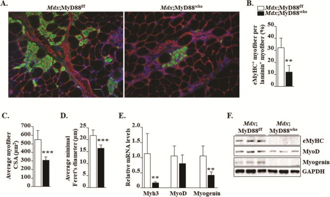
Figure 4.

Ablation of MyD88 in satellite cells inhibits myofiber regeneration in mdx mice. (A) Representative photomicrographs of transverse GA muscle sections of Mdx;MyD88f/f and Mdx;MyD88scko mice immunostained for eMyHC (green color) and laminin (red color). Nuclei stained with DAPI. (B) Percentage of eMyHC+ myofiber per laminin+ myofiber. Quantification of eMyHC+ myofiber (C) CSA and (D) minimal Feret’s diameter in GA muscle of Mdx;MyD88f/f and Mdx;MyD88scko mice. N = 6 or 7 in each group. (E) Relative mRNA levels of regeneration markers Myh3, MyoD and myogenin in GA muscle of Mdx;MyD88f/f and Mdx;MyD88scko mice. (F) Representative immunoblots showing levels of eMyHC, MyoD and myogenin and an unrelated protein GAPDH in GA muscle of Mdx;MyD88f/f and Mdx;MyD88scko mice. Scale bar: 50 μm. N = 5 mice in each group. Error bars represent s.d. *P-value < 0.05, **P-value < 0.01 and ***P-value < 0.001 from corresponding littermate Mdx;MyD88f/f mice by unpaired t-test.
