
Figure 5.
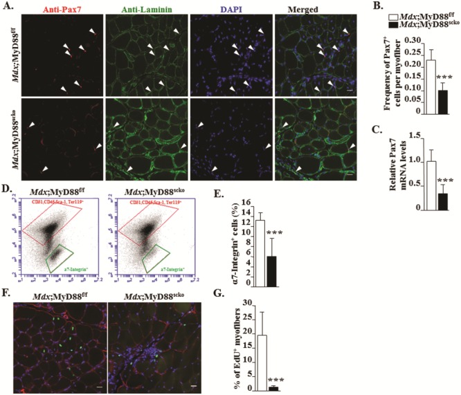
Ablation of MyD88 reduces satellite cell number and their fusion with injured myofibers in mdx mice. (A) Representative photomicrographs of transverse sections of GA muscle of Mdx;MyD88f/f and Mdx;MyD88scko mice after immunostaining for Pax7 (red) and laminin (green). Nuclei were labelled with DAPI (blue). (B) Quantitative analysis showing frequency of Pax7+ cells per myofiber in Mdx;MyD88f/f and Mdx;MyD88scko mice. (C) Relative mRNA levels of Pax7 in GA muscle. (D) Representative FACS dot plots showing the percentage of satellite cells in GA muscle of Mdx;MyD88f/f and Mdx;MyD88scko mice. Negative selection antibodies (CD31, CD45, Ter119) are in upper box, whereas positive selection antibody (α7-Integrin) is in lower box. (E) Quantification of percentage of α7-Integrin+ cells in GA muscle of Mdx;MyD88f/f and Mdx;MyD88scko mice. N = 5 in each group. (F)Mdx;MyD88f/f and Mdx;MyD88scko mice were processed for the detection of EdU+ nuclei (green colored). The sections were also stained for laminin (red colored). Nuclei were counterstained with DAPI. (G) Percentage of EdU+ nuclei per myofiber. Scale bar: 20 μm. N = 5 for Mdx;MyD88f/f and N = 7 for Mdx;MyD88scko group. Error bars represent s.d. *P-value < 0.05, **P-value < 0.01 and ***P-value < 0.001 from corresponding littermate Mdx;MyD88f/f mice by unpaired t-test.