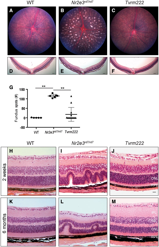
Figure 1.
Tvrm222 mice show suppression of rd7-associated photoreceptor dysplasia. Fundus images (A–C; quantification of the numbers of dysplastic lesions observed in fundus images of the central retina are shown in G) and posterior eye sections stained with H&E (D–F) from 1-month-old WT, Nr2e3rd7/rd7, and Nr2e3rd7/rd7; Frmd4bTvrm222/Tvrm222 (Tvrm222) mice. Note spotting phenotype in the rd7 fundus image (B) and the corresponding retinal folds or lesions shown in histological sections (E). Scale bar=100μm. For quantification in (G), the results are mean±SD, n = 5 (WT), 6 (rd7) or 13 (Tvrm222); **P < 0.0001 (one-way ANOVA, post-hoc Tukey’s test). (H-M) Representative retinal sections from 2-week-old (H–J) and 6-month-old (K–M) WT, Nr2e3rd7/rd7 and Tvrm222 mice are shown by H&E staining. Note the undulation of the retinal layers in the Nr2e3rd7/rd7 mice and the absence of retinal folds in sections from Tvrm222 mice at both ages surveyed. Scale bar=20μm. IPL, inner plexiform layer; INL, inner nuclear layer; ONL, outer nuclear layer; RPE, retinal pigment epithelium.

