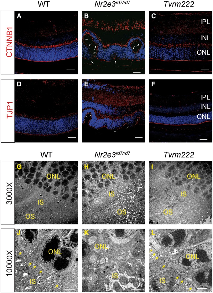
Figure 3.
Suppression of photoreceptor dysplasia in Tvrm222 mice is associated with normalization of retinal lamination, and intact cell junctions at the ELM. (A–F) Retinal sections from 1-month-old WT, Nr2e3rd7/rd7 and Tvrm222 mice were subject to immunofluorescence staining using antibodies against CTNNB1 (β-catenin) (A–C) and TJP1 (ZO-1) (D–F), respectively. Scale bar=20 μm. The fragmentation of the ELM is marked by arrows. Ultra-structural assessment of the retinal architecture from 1-month-old WT (G, J), Nr2e3rd7/rd7 (H, K) and Tvrm222 (I, L) mice. The ELM is marked by yellow arrows. Scale bar=5 μm (×3000) or 2 μm (×10 000). IPL, inner plexiform layer; INL, inner nuclear layer; ONL, outer nuclear layer; IS, inner segment; OS, outer segment.

