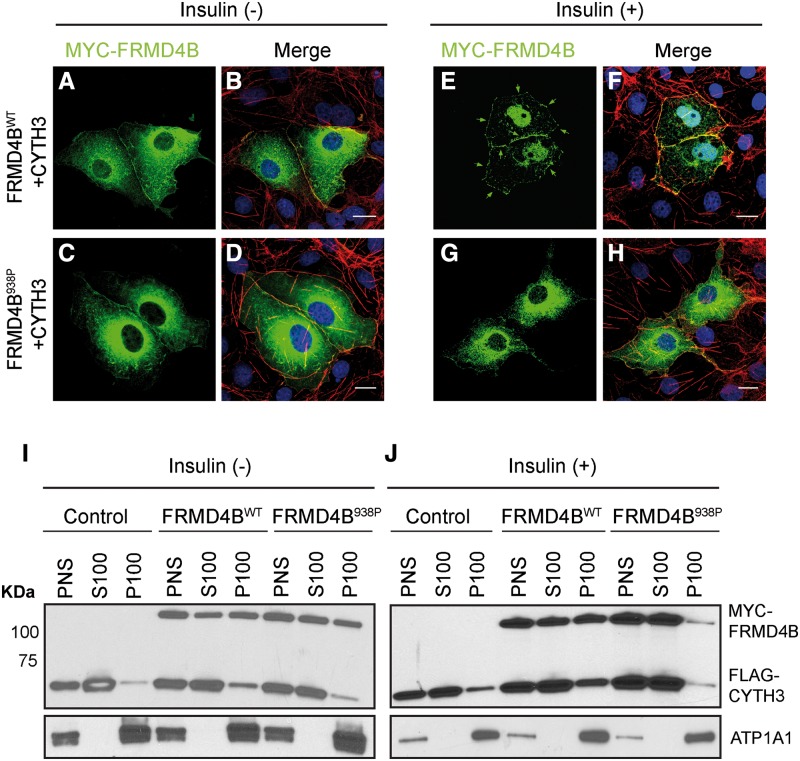
Figure 6.
Membrane recruitment of FRMD4B938P upon insulin stimulation is reduced in vitro (A–H). COS7 cells were co-transfected with 3×FLAG-CYTH3 and either 3×myc-FRMD4Bwt (A, B; E, F) or 3×myc-FRMD4B938P (C, D; G, H), and subject to serum starvation (without insulin; A–D) or serum starvation followed by treatment with 100 nM insulin (E–H). 3×myc-FRMD4B was detected by immunofluorescence using antibody against the myc tag (green). Counterstaining with phalloidin (red) shows sub-plasma membrane actin cytoskeleton. Note the cell surface associated FRMD4Bwt (arrows). Scale bar=20 μm. (I, J) Subcellular fractionation profiles of HEK-293T cells transiently co-transfected with 3×FLAG-CYTH3 and either 3×myc-FRMD4Bwt or 3×myc-FRMD4B938P, and untreated (I) or treated (J) with insulin after serum starvation. Na+/K+-ATPase (ATP1A1) was used as the loading control for membrane fractions. PNS, post-nuclear supernatant; S100, supernatant after 100 000×g centrifugation; P100, pellet after 100 000×g centrifugation.

