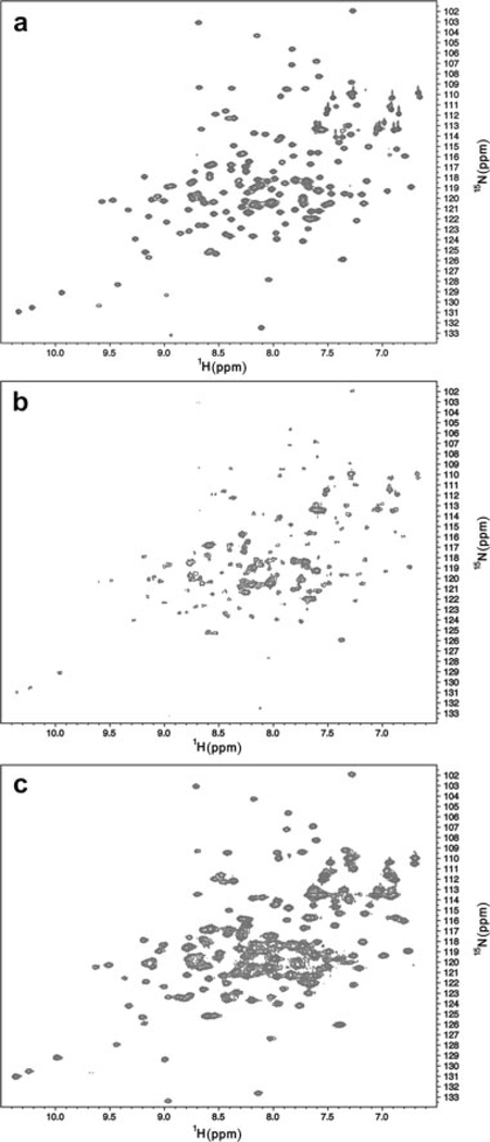
Figure 3.
15N HSQC spectra of WT* T4 lysozyme in solution. (a) The spectrum of the protein in MOPS buffer containing 50mM NaCl in the absence of adjuvant. (b) The spectrum obtained in the same MOPS buffer containing 50mM NaCl in the presence of adjuvant. The reduction in signal intensity is due to the binding of protein to adjuvant. (c) The spectrum obtained in the presence of the same MOPS buffer in 200mM NaCl and adjuvant. Recovery of the signal is due to the desorption of protein from adjuvant. The spectrum suggests minimal perturbation to the protein as a result of binding.

