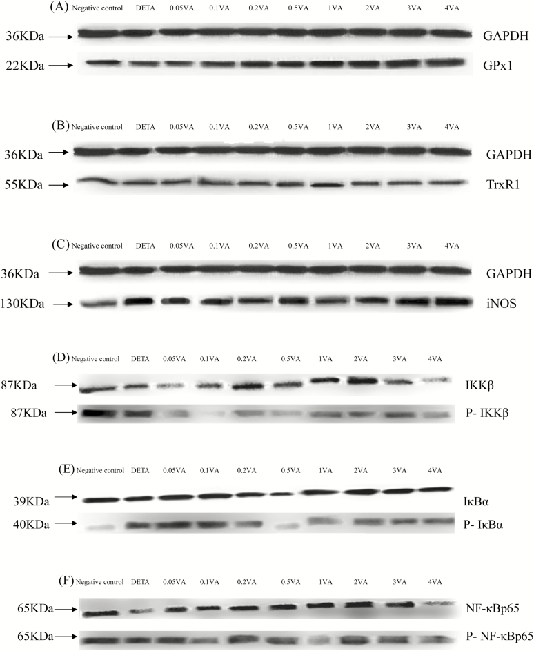
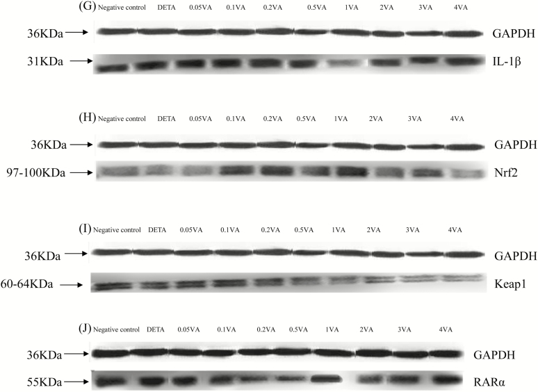
Figure 3.
Effect of VA on DETA-induced selenoproteins, RARα, IL-1β protein expression, and phosphorylation of Nrf2 and NF-κB pathways in BMEC. The cells were randomly divided into 10 groups with 6 replicates, and 3 independent experiments were performed. The first group was used as negative control: without VA (Sigma-Aldrich, Munich, Germany) and DETA-NO (Sigma-Aldrich, Munich, Germany) for 30 h. Group 2 was DETA-NO-treated group: without VA for 24 h before treated with DETA-NO (1,000 μmol/liter) for an additional 6 h. Groups 3–10 were 8 doses of VA plus DETA-NO-treated groups: pretreated BMEC with 0.05, 0.1, 0.2, 0.5, 1, 2, 3, or 4 μg/mL of VA for 24 h and then incubated in the presence of 1,000 μmol/liter of DETA-NO and VA for a further 6 h. Expressions of GPx1 (A), TrxR1 (B), iNOS (C), IL-1β (G), RARα (J), Nrf2 (H), Keap1 (I), and phosphorylated IKKβ (D), IκBα (E), NF-κBp65 (F) protein levels were detected by western blotting and normalized to GAPDH levels.


