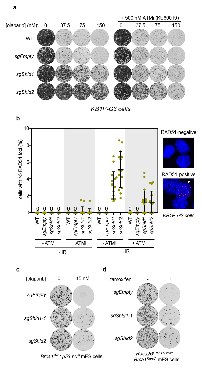
ED Figure 3. Supporting data that mouse Shieldin promotes resistance to PARP inhibition in Brca1-mutated cells and tumours.
a, Clonogenic survival assays of transduced KB1P-G3 cells treated with indicated olaparib doses ± ATM inhibitor (ATMi) KU60019 (500 nM). On day 6, the ATMi alone and untreated groups were stopped and stained with 0.1% crystal violet; the other groups were stopped and stained on day 9. Data shown is a representative set from 3 biologically independent experiments (with 3 technical replicates each). b, Left, quantitation of Rad51 focus formation in parental KB1P-G3 (Brca1-/-; Trp53-/-) cells or KB1P-G3 cells that were transduced with the indicated lentiviral sgRNA vectors. Cells were fixed without treatment or 4 h after irradiation (10 Gy dose). Each data point represents a microscopy field containing a minimum of 50 cells; the bar represents the mean ± SD (n=15). Right, representative micrographs of Rad51-negative and -positive cells (the latter is indicated by an arrowhead). DNA was stained with DAPI. c, Clonogenic survival assay of Rosa26CreERT2/wt; Brca1Δ/Δ; p53-null mES-cells virally transduced with the indicated sgRNA and treated without or with 15 nM olaparib for 7 d. Gene editing efficiencies of the sgRNAs can be found in Supplementary Table 2. Data shown is a representative set from 3 biologically independent experiments (with ≥ 2 technical replicates each). d, Clonogenic survival assay of Rosa26CreERT2/wt;Brca1Sco/Δ mES-cells virally transduced with the indicated sgRNA and treated without or with 0.5 µM tamoxifen to induce BRCA1 depletion. Gene editing efficiencies of the sgRNAs can be found in Supplementary Table 2. Data shown is a representative set from 2 biologically independent experiments (with 3 technical replicates each).

