FIGURE 2.

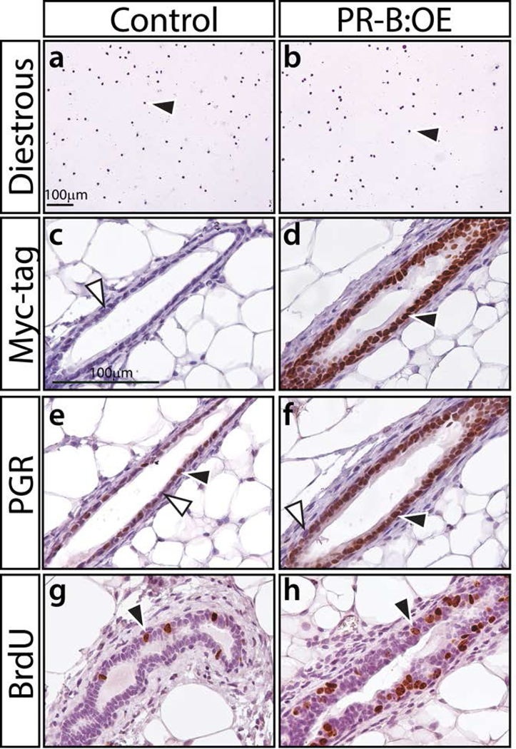
The PGR-B isoform is specifically targeted to the mammary epithelium of the PR-B:OE mouse. (a) and (b) show crystal violet stained vaginal cytology from 8-week old control and PR-B:OE virgin mice respectively. Both genotypes were in diestrus at the time of euthanasia; black arrowhead indicates the presence of leucocytes, which are indicative of the diestrus stage of the estrous cycle. (c) and (d) show a transverse section of a mammary gland duct from control and PR-B:OE mice respectively, which has been stained for myc-tag immunopositivity. Note the expected absence of positive staining in the control gland (white arrowhead) whereas the PR-B:OE mammary gland duct shows strong staining for the myc-epitope (black arrowhead), which is localized to the epithelial compartment of the mammary duct. (e) and (f) display mammary ducts from control and PR-B:OE mice stained for PGR expression respectively. Note control mammary gland duct shows the typical punctate spatial pattern for PGR expression within the epithelial compartment (positive and negative staining indicated by black and white arrowheads respectively). In contrast, the PR-B:OE mammary gland duct shows stronger PGR staining for the majority of the mammary epithelial cells. (g) and (h) show typical BrdU staining results for the control and PR-B:OE mammary gland respectively at diestrus. Note a significant increase in the number of epithelial cells that score positive for BrdU incorporation (black arrowhead) in the PR-B:OE gland compared to the control gland.
