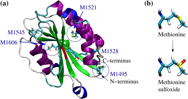
Fig 1. Crystallographic structure of the A2 domain and oxidation of methionine residues.
(a) Cartoon representation of the A2 domain (PDB code 3GXB). The backbone is colored according to secondary structure elements with α helices in purple, 310 helices in blue, β strands in green and turns in cyan. The proteolysis site is highlighted in red. The five methionine residues contained within the core of the A2 domain are shown in the stick and ball representation. Blue circles indicate three methionine residues that were determined to be oxidized in mass spectrometry measurements in the presence of HOCl [5]. The other two methionine residues are also likely to be oxidized under the same conditions. (b) Conversion pathway from methionine to methionine sulfoxide.

