
Figure 2.
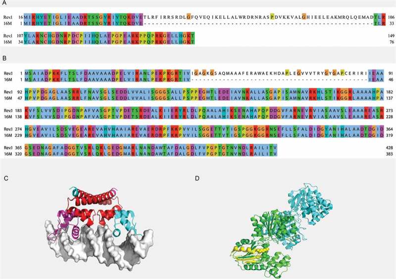
Regions of insertions within the MerR (a) and BMEI1570 (b) proteins of the B. melitensis Rev.1 strain. Pairwise alignment between the Rev.1 (top) and 16M (bottom) sequences was visualized using the Clustal X color scheme for residues coloring [63]. (c) Crystal structure of the E. Coli copper efflux regulator homodimer (4WLS). DNA is shown as white surface. The inserted sequence 1 (RI1) is shown in red. (d) Crystal structure of the T. maritima glycerate kinase (2B8N). The inserted sequence 2 (RI2) is shown in yellow. Active site residues are shown as spheres.