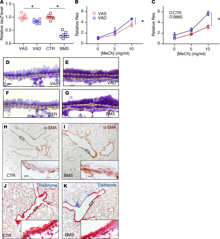
Figure 1. Disruption of RA signaling in vivo for 5 days results in airway hyperresponsiveness and airway remodeling.
(A) RA activity is downregulated in RA-deficient lung as evidenced by the reduction of lacZ expression in VAD (compared with VAS) and BMS (compared with CTR) in RARE-lacZ lungs (n = 6 per group). (B and C) Airway resistance (Res) is higher in the VAD (B) and BMS (C) mice compared with VAS (B) and CTR (C) mice when challenged with increasing doses of the bronchoconstrictor methacholine (MeCh), indicating higher airway responsiveness in RA-deficient animals (n = 9 per group). (D–G) Plastic sections of mouse lungs showing thicker ASM layer (delineated by yellow dotted lines) in VAD (E) and BMS (G) airways compared with VAS (D) and CTR (F) airways (n = 3 per group). Epi labels the airway epithelial layer. (H and I) Immunostaining of smooth muscle–specific marker α-SMA showing an increase in ASM mass in BMS (I) airway compared with the CTR (H) airway (n = 3 per group). (J and K) Trichrome staining revealing increased collagen content (stained blue) in the BMS (K) airway compared to CTR (J) airway (n = 3 per group). Data represent the mean ± SEM (A) or mean ± range (B and C). Statistical significance was calculated using Student’s t test (A) and 2-way ANOVA (B and C). *P < 0.05. Scale bars: 10 μm (D, H, and J).

