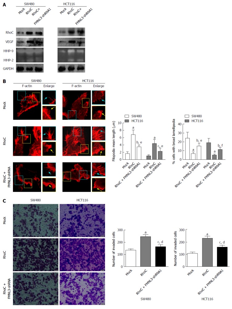
Figure 4.

Formin-like 3 depletion blocks RhoC-dependent increases of matrix metalloproteins and vascular endothelial growth factor (A), assembly of actin-based protrusions (B), and invasion (C) in colorectal carcinoma cells. MMPs and VEGF were detected by gelatin zymography experiments and western blot. F-actin is displayed using tritc-phalloidine (Red) staining and laser scanning confocal microscopy detection. Enlarged views of the boxed regions are shown on the right side of the figures. Blue arrows indicate filopodia, yellow arrows indicate lamellipodia. Cell invasion was compared using the Boyden chamber assay. Scale bars represent 5 μm (F-actin) or 50 μm (cell invasion assay), respectively. aP < 0.001, bP < 0.01 and cP < 0.05 vs Mock group, dP < 0.001, eP < 0.01 and fP < 0.05 vs RhoC-overexpressing group. Error bars indicate mean ± SD. FMNL3: Formin-like 3; MMP: Matrix metalloprotein; VEGF: Vascular endothelial growth factor.
