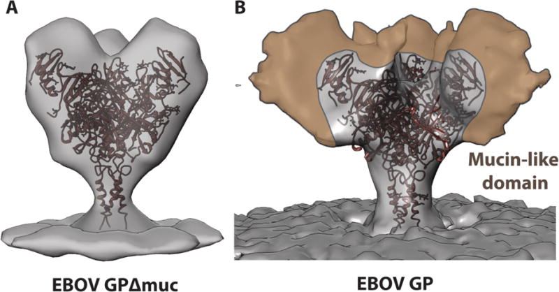
Figure 2. Visualization of the Mucin-Like Domain.

(A) The crystal structure of the mucin-deleted EBOV GP (PDB: 5JQ3) [65] is shown docked into a subtomogram averaged map of mucin-deleted EBOV GP [66] and a single-particle generated map of intact, mucin-containing EBOV GP (B) [22]. Although the observation of density for mobile regions is limited by technical factors in single particle reconstruction, the regions of the mucin-like domain that are visible appear to extend upwards and outwards from the glycan cap and base region, thereby shielding much of the GP core.
