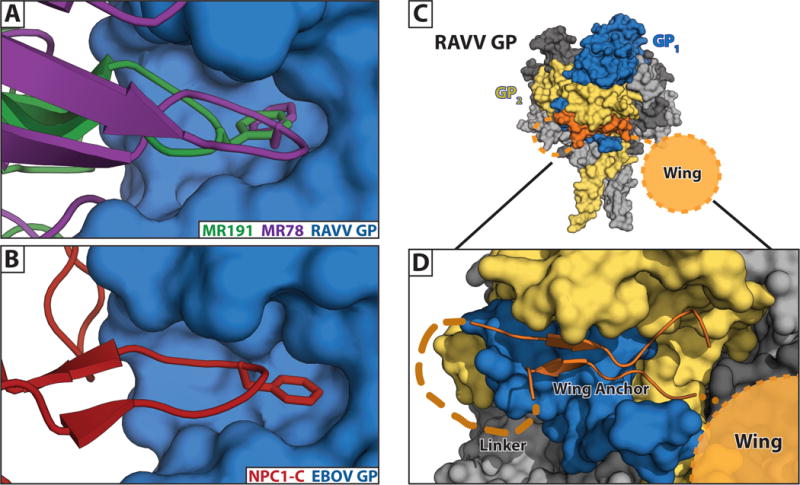
Figure 4. Neutralizing epitopes identified for marburgvirus.

(A) Crystal structures of neutralizing antibodies MR191 [25] and MR78 [49] bound within the RBS of RAVV GP. (B) A phenylalanine at the apex of the CDR-H3s of both antibodies reaches into the hydrophobic pocket of the marburgvirus GP in a manner that structurally mimics interactions of ebolavirus GP both with its glycan cap and its host receptor, NPC1-C [12]. (C) A marburgvirus GP is shown with a single GP monomer colored in blue (GP1) and gold (GP2). The remaining two monomers are grey. The anchor of the wing domain (orange) is shown wedged underneath the base of GP. (D) Enlarged view of the wing illustrating the β-strand wing anchor region (connected by an 8 aa linker), and the relative position of the 33 aa wing targeted by antibodies.
