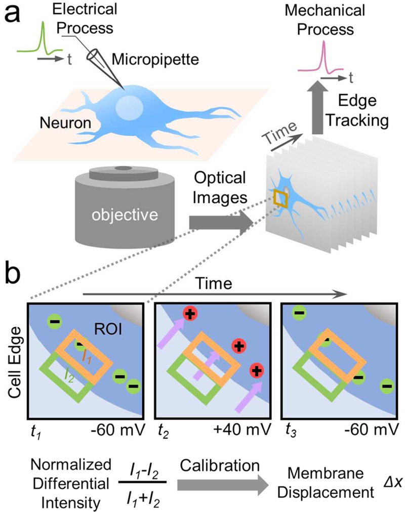
Figure 1: Optical imaging of mechanical motion accompanying action potential in single mammalian neurons.

a) Experimental setup showing a hippocampal neuron cultured on a glass slide mounted on an inverted optical microscope, which is studied simultaneously with the patch clamp configuration for electrical recording of the action potential, and optical imaging of membrane displacement associated with the action potential. b) Imaging and quantification of membrane displacement with a differential detection algorithm that tracks the edge movement of a neuron. A region of interest is selected to include a portion of the neuron edge, which is divided into two halves along the edge (orange and green boxes) with intensities, I1 and I2, respectively. The expansion or shrinkage of the neuron is measured by a membrane displacement,Δx, from the change in the intensities, which is proportional to the normalized differential intensity, (I1-I2)/(I1+I2). See text for details.
