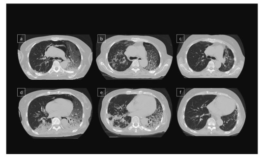
Fig. 5.

Time course of chest CT images.
(a,d) Pneumomediastinum, consolidation and patchy GGO were shown in both lungs. (b,e) Pneumomediastinum was disappeared 2 months after immunosuppressive treatment. GGO and consolidation were still showed bilateral lobes.
(c,f) CT showed lung fibrosis mainly in the left lobe, but consolidation and GGO dramatically improved 1 year after immunosuppressive treatment.
