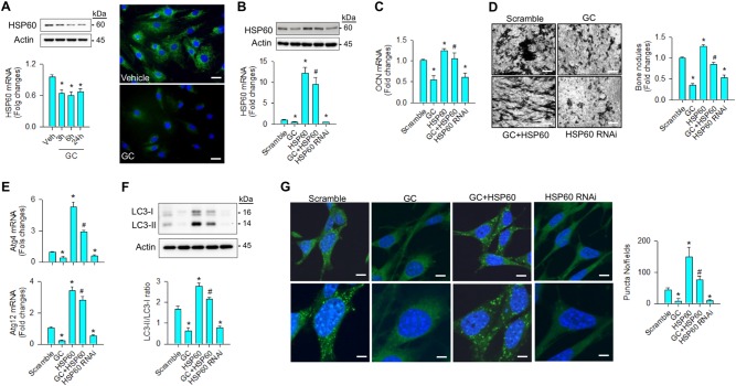
Fig. 1. Analyses of HSP60-mediated autophagy and differentiation of osteoblasts.
a Glucocorticoid reduced mRNA expression, protein levels, and immunofluorescence of HSP60 in osteoblasts. Scale bar, 8 μm. b Forced HSP60 expression attenuated the glucocorticoid-induced loss of (c) osteocalcin expression and d mineralized matrix accumulation. Scale bar, 40 μm. Increasing HSP60 attenuated the glucocorticoid-mediated loss of (e) Atg4, and Atg12 expressions, (f) LC3-II levels, LC3-II/LC3-I ratio, and g autophagic puncta formation. Scale bars, 6 μm (upper panels) and 12 μm (lower panels). Knocking down HSP60 decreased autophagic marker expressions, autophagic vesicle formation, and mineralized matrix production of osteoblasts. Experiments results are expressed as mean ± standard error. Asterisks (*) resemble a significant difference (P < 0.05) vs. vehicle or scramble group, and hashtag (#) indicates a distinguishable difference (P < 0.05) vs. glucocorticoid-treated group. Veh, vehicle; GC, glucocorticoid

