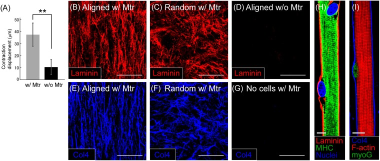
Figure 3.
(A) Comparison of contraction displacement between myofiber sheets cultured in the fibrin-based gels with (w/ Mtr) and without Matrigel (w/o Mtr). (n = 5) (**P < 0.01) (B,C) Laminin structure shown in the gel containing Matrigel (w/ Mtr). Myofibers are aligned (B) or randomly oriented in the gel (C). (D) No laminin formation observed in the gel without Matrigel (w/o Mtr). The aligned cells produced no laminin structure in the gel. (E,F) Formation of anisotropic and isotropic structures of type IV collagen in the Matrigel-containing gels with (E) aligned and (F) randomly oriented myofibers. (G) No formation of type IV collagen in the gel containing Matrigel, but without cells. (H) Laminin surrounding a myofiber in the myofiber sheet. (red: laminin, green: myosin heavy chain (MHC), blue: nuclei) (I) Type IV collagen surrounding a myofiber (blue: Type IV collagen, red: F-actin, green: myogenin (myoG)) Scale bars: 100 μm (B–G), 10 μm (H,I).

