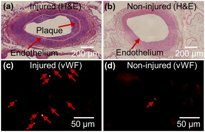
Figure 5.
Haematoxylin and eosin stained vessel on the injured side (a) and non-injured side (b). Thirty images acquired at ×40 magnification are stitched to reconstruct an overall image of the vessel for (a) and (b). Significant plaque development is found in the injured side (a). Vasa vasorum on adventitia in the selected region was stained by anti-von Willebrand factor. A large number of vasa vasorums are found in adventitia on the injured side (c), but a few vasa vasorums are found in adventitia on the non-injured side (d).

