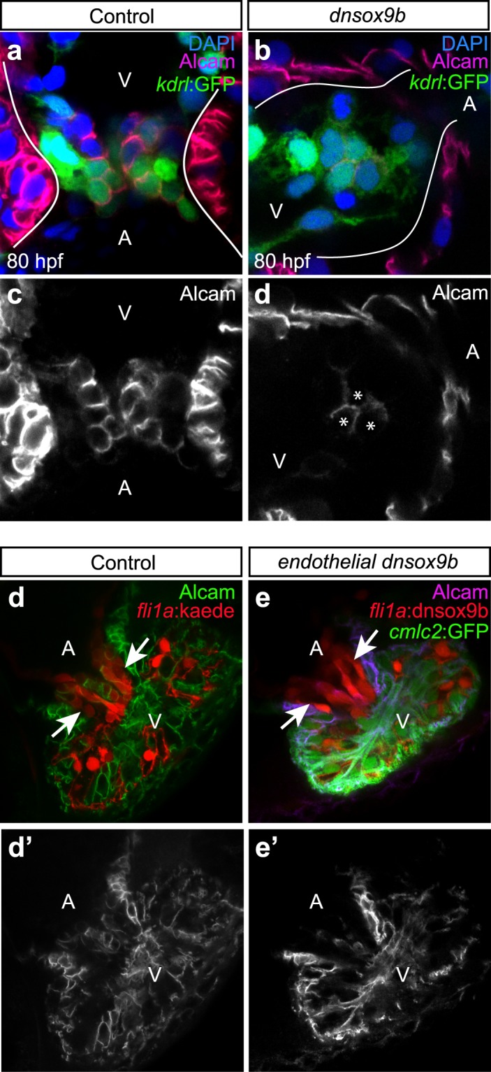
Figure 4.

Cardiomyocyte-specific but not endothelial-specific inhibition of sox9b function results in hypoplastic atrioventricular cushions. Control larvae (a,c,d,d’; Tg(myl7:Gal4VP16;kdrl:GFP)) and larvae with cardiomyocyte-specific loss of sox9b function (b,d,e,e’; Tg(myl7:Gal4VP16;kdrl:GFP;UAS:dnsox9b-2A-tRFP)) were fixed at 80 hpf and processed for fluorescent immunohistochemistry using antibodies against activated leukocyte cell adhesion molecule (Alcam; purple). (a,b) Larvae were mounted ventrally in low melting point agarose and imaged at 40x magnification with a confocal microscope. (c,d) Boxed areas in a & b showing endocardial expressing Alcam, a marker of differentiated endocardial cushion cells. (a,c) AV cushions form normally in control larvae, as indicated by a coalescence of Alcam + endocardial cells at the junction between the atrium and ventricle (boxed area in a). (b,d) Larvae with in cardiomyocyte-specific inhibition of sox9b had hypoplastic cushions with Alcam + endocardial cells. White arrow indicates endocardium pushing through a hole in myocardium. Ventricle (V) and atrium (At) are abbreviated as indicated. Images are representative phenotypes, n = 5 per group. (e-f’) Control larvae (Tg(fli1a:Gal4ff;UAS:Kaede)) and larvae with endothelial-specific loss of sox9b function Tg(fli1a:Gal4ff;UAS:dnsox9b-2A-tRFP; UAS:Kaede)) were fixed at 120 hpf, processed for fluorescent immunohistochemistry using antibodies against Alcam, and scored for the presence of endothelial cushion. Endothelial cushions clearly formed in both control larvae (e) and larvae with endothelial-specific loss of sox9b function (f). Images are representative phenotypes, n = 8 per group.
